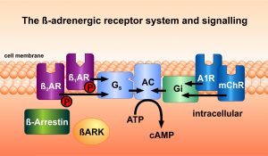
Adrenergic receptors play a pivotal role in the regulation of many physiological functions of the cardiovascular system. Catecholamines such as adrenaline and norepinephrine bind to these receptors which are common in many organs of the body. These receptors are activated by specific ligands, and can then be specifically inactivated by proteins which belong to the families of receptor kinases and of arrestins. The team has developed a number of compounds which bind to these proteins, and regulate the propagation of hormone stimuli to intracellular signalling pathways.
Additionally, the group of W. Koch and the 2012 Nobel prize awardee Robert J. Lefkowitz, in whose group Martin Lohse had worked for several years, investigated gene transfer of the C-terminal fragment „ßARKct“ of the ß-adrenergic receptor kinase-1 (ßARK-1, GRK-2) for its effect on heart muscle cells. They observed an improvement of myocardial function in heart failure in several animal models. Our team has extended these observations to an in vivo gene transfer model and to a related signalling protein, phosducin. New insight into the intracellular compartmentation of second messengers, especially cAMP, was obtained in cooperations. Methods to detect this signal activation and signal spreading optically on a millisecond scale were developed.
Specifically, novel assays were established which allow to determine molecular arrestin activation. Thereby, signal pathway-specific receptor agonists (“biased ligands”) can be identified.
References on receptor pharmacology and signalling
- Anton SE, Kayser C, Maiellaro I, Nemec K, Möller J, Koschinski A, Zaccolo M, Annibale P, Falcke M, Lohse MJ, Bock A. Receptor-associated
independent cAMP nanodomains mediate spatiotemporal specificity of GPCR signaling. Cell 2022;185(7):1130-1142.e11. doi: 10.1016/j.cell.2022.02.011 - Bathe-Peters M, Gmach P, Boltz HH, Einsiedel J, Gotthardt M, Hübner H, Gmeiner P, Lohse MJ, Annibale P. Visualization of β-adrenergic
receptor dynamics and differential localization in cardiomyocytes. Proc Natl Acad Sci U S A. 2021;118(23): e2101119118. doi: 10.1073/pnas.2101119118 - Bock A, Annibale P, Konrad C, Hannawacker A, Anton SE, Maiellaro I, Zabel U, Sivaramakrishnan S, Falcke M, Lohse MJ. Optical Mapping of cAMP
Signaling at the Nanometer Scale. Cell 2020; 182: 1519–1530; e1–e17; doi: 10.1016/j.cell.2020.07.035 - Möller J, Isbilir A, Sungkaworn T, Osberg B, Karathanasis C, Sunkara V, Grushevskyi EO, Bock A, Annibale P, Heilemann M, Schütte C, Lohse
MJ. Single-molecule analysis reveals agonist-specific dimer formation of µ-opioid receptors. Nat Chem Biol 2020;16(9):946-954. doi:
10.1038/s41589-020-0566-1 - Annibale P, Lohse MJ. Spatial heterogeneity in molecular brightness. Nature Methods 2020;17(3):273-275. doi: 10.1038/s41592-020-0732-0
- Işbilir A, Möller J, Arimont M, Bobkov V, Perpiñá-Viciano C, Hoffmann C, Inoue A, Heukers R, de Graaf C, Smit MJ, Annibale P, Lohse
MJ. Advanced fluorescence microscopy reveals disruption of dynamic CXCR4 dimerization by subpocket-specific inverse agonists. Proc Natl Acad Sci US A 2020;117(46):29144-29154. doi: 10.1073/pnas.2013319117 - Grushevskyi EO, Kukaj T, Schmauder R, Bock A, Zabel U, Schwabe T, Benndorf K, Lohse MJ. Stepwise activation of a class C GPCR begins with
millisecond dimer rearrangement. Proc Natl Acad Sci U S A 2019; 116(20):10150-10155. doi: 10.1073/pnas.1900261116. - Sungkaworn T, Jobin ML, Burnecki K, Weron A, Lohse MJ, Calebiro D. Single-molecule imaging reveals receptor-G protein interactions at cell
surface hot spots. Nature 2017; 550(7677):543-547. doi: 10.1038/nature24264 - Godbole A, Lyga S, Lohse MJ, Calebiro D. Internalized TSH receptors en route to the TGN induce local Gs-protein signaling and gene transcription. Nature Commun. 2017;8(1):443
- Emami-Nemini A, Roux T, Leblay M, Bourrier E, Lamarque L, Trinquet E, Lohse MJ Time-resolved fluorescence ligand binding for G-protein-coupled receptors. Nature Prot 2013; 8: 1307-1320.
- Lohse MJ, Calebiro D. Receptor signals come in waves. Nature 2013; 495: 457-458.
- Calebiro D, Rieken F, Wagner J, Sungkaworn T, Zabel U, Borzi A, Cocucci E, Zürn A, Lohse MJ. Single-molecule analysis of fluorescently
labeled GPCRs reveals receptor-specific complexes with distinct dynamics and organization. Proc. Natl. Acad. Sci. USA 2013; 110:
743–748. - Hlavackova V, Zabel U, Frankova D, Bätz J, Hoffmann C, Prezeau L, Pin J-P, Blahos J, Lohse MJ. Sequential inter- and intra-subunit rearrangements during asymmetric activation of dimeric metabotropic glutamate receptor 1. Science Sign 2012; 5: ra59.
- Lohse MJ, Nuber S, Hofmann C. Fluorescence/bioluminescence resonance energy transfer techniques to study G-protein-coupled receptor
activation and signalling. Pharm Rev. 2012; 64: 299–336. - Calebiro D, Nikolaev VO, Gagliani MC, de Filippis T, Dees C, Tacchetti C, Persani L, Lohse MJ Persistent cAMP-signals triggered by internalized G-protein-coupled receptors. PLoS Biol 2009; 7:e1000172.
- Vilardaga JP, Nikolaev VO, Lorenz K, Ferrandon S, Zhuang Z, Lohse MJ. Conformational cross-talk between alpha 2A-adrenergic and mu-opioid
receptors controls cell signaling. Nature Chemical Biology 2008; 4: 126-131 - Lorenz K, Schmitt JP, Schmitteckert EM, Lohse MJ. A new type of ERK1/2-autophosphorylation causes cardiac hypertrophy. Nature Medicine 2009; 15: 75-83
- Nikolaev VO, Bünemann M, Schmitteckert E, Lohse MJ, Engelhardt S. Cyclic AMP imaging in adult cardiac myocytes reveals far-reaching ß1-adrenergic but locally confined alpha2-adrenergic receptor-mediated signaling. Circulation Research 2006; 99: 1084-1091.
- Buitrago M, Lorenz K, Maass AH, Oberdorf-Maass S, Keller U, Schmitteckert EM, Ivashchenko Y, Lohse MJ, Engelhardt S. The transcriptional repressor Nab1 is a specific regulator of pathological cardiac hypertrophy. Nature Medicine 2005; 11: 837-844.
- Hein P, Frank M, Hoffmann C, Lohse MJ, Bünemann M. Dynamics of receptor/G protein coupling in living cells. EMBO J. 2005; 24: 4106-4114.
- Hoffmann C, Gaietta G, Bünemann M, Adams S, Oberdorff-Maass S, Behr B, Vilardaga JP, Tsien RY, Ellisman MH, Lohse MJ. A FlAsH-based FRET
approach to determine G-protein coupled receptor activation in living cells. Nature Methods 2005; 2: 171-176. - Vilardaga JP, Steinmeyer R, Harms GS, Lohse MJ. Molecular basis of inverse agonism in a G protein-coupled receptor. Nature Chem. Biol. 2005; 1: 25-28.
- Nikolaev VO, Bünemann M, Hein L, Hannawacker A, Lohse MJ. Novel single chain cAMP sensors for receptor-induced signal propagation. J. Biol. Chem 2004; 279: 37215-37218.
- Bünemann M, Frank M, Lohse MJ. Gi protein activation in intact cells involves subunit rearrangement rather than dissociation. Proc. Natl. Acad. Sci. USA 2003; 100: 16077-16082.
- Engelhardt S, Hein L, Dyachenkov V, Kranias EG, Isenberg G, Lohse MJ. Altered calcium handling is critically involved in the cardiotoxic effects of chronic ß-adrenergic stimulation. Circulation 2004; 109: 1154-1160
- Li Z, Laugwitz KL, Pinkernell K, Pragst I, Baumgartner C, Hoffmann E, Rosport K, Münch G, Humrich J, Lohse MJ, Ungerer M. Effects
of two Gbetagamma-binding proteins–N-terminally truncated phosducin and beta-adrenergic receptor kinase C terminus (ßARKct)–in heart failure. Gene Therapy 2003;10(16):1354-61 - Lorenz K, Lohse MJ, Quitterer U. Protein kinase C switches the Raf kinase inhibitor from Raf-1 to GRK-2. Nature 2003; 426: 574-579
- Vilardaga JP, Bünemann M, Krasel C, Castro M, Lohse MJ. Measurement of the millisecond activation switch of G-protein-coupled receptors in
living cells. Nature Biotechnology 2003; 21: 807-812. - Ungerer M, Kessebohm K, Kronsbein K, Lohse MJ, Richardt G. Activation of ß-adrenergic receptor kinase during myocardial ischemia. Circulation Research 1996; 79: 455-460
- Ungerer M, Parruti G, Böhm M, Puzicha M, DeBlasi A, Erdmann E, Lohse MJ. Expression of ß-arrestins and ß-adrenergic receptor kinases in the
failing human heart. Circulation Research 1994; 74:206-213 - Ungerer M, Böhm M, Elce JS, Erdmann E, Lohse MJ. Altered expression of ß-adrenergic receptor kinase (ßARK) and ß1-adrenergic receptors in
the failing human heart. Circulation 1993; 87:454-463 - Bauer PH, Müller S, Puzicha M, Pippig S, Obermaier B, Helmreich EJ, Lohse MJ. Phosducin is a protein kinase A-regulated G-protein regulator.
Nature 1992; 358(6381): 73-76 - Lohse MJ, Benovic J, Codina J, Caron MG, Lefkowitz RJ. ß-Arrestin: a protein that regulates ß-adrenergic receptor function. Science 1990;248: 1547-1550